Tomography of the Atlas vertebra

3D Spiral CT - Cone Beam CT
Since time immemorial, people have tried to find ways of looking inside the human body to understand how it works and to detect dysfunctions without having to resort to invasive methods.Each imaging method has its own peculiarities, limitations, advantages and disadvantages. Depending on what is to be visualised, one method may be more suitable than another. Since this is an important technical health topic, which usually confuses ordinary people who are not familiar with the subject, we prefer to deal with it in detail.
Foreword
Expecting a doctor or a radiologist to tell you about the misalignment of the Atlas is equivalent to expecting a painter to be an expert on Picasso's works. They both paint, but in completely different ways. You may be lucky enough that the painter not only paints walls, but is also an art lover and therefore an expert on Picasso, but in most cases you will only make a dead end. This concept must be very clear to you.Do not expect to find anything in your radiology reports about the misalignment of your Atlas. Only recently, and only after having devised a method and the necessary equipment to effectively and stably correct the first cervical vertebra, has it been possible to identify the Atlas as a possible cause of certain complaints. In conventional medical books, the misaligned Atlas does not appear as a pathological condition, so that no investigations are carried out in this direction by doctors and radiologists. As a result, the misaligned Atlas is very rarely found in a radiological report.
If you want to inform yourself and make use of a new technology, you cannot ask someone who still applies the old one, regardless of their training! You cannot ask someone who drives a petrol car what it is like to drive an electric Tesla.
It is a common misconception that a doctor has to know everything, just because he is a doctor and deals with health. New discoveries are made all the time that your doctor may not be aware of.
If you want competent and detailed information about the Atlas and its stable correction, you cannot ask your doctor or therapist, who have no experience in this area. What little they can tell you is not necessarily true. Understanding this reality will greatly benefit your health.
Atlantomed is your point of reference when it comes to high cervical misalignment.
Diagnostic imaging methods
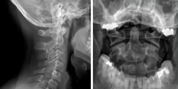
Radiography

The normal X-ray that we all know works like this: a cathode ray tube generates X-rays that are passed through the body and captured by a sensor on the opposite side. Because the bones are much denser than the soft parts, they allow fewer rays to pass through and as a result, the X-ray image is white, while the soft parts are darker.
With the development of technology and the increased sensitivity of sensors, very small doses of X-rays are now needed to obtain an X-ray image. You can find a detailed explanation of ionising radiation here: Ionising radiation and cancer.
An X-ray image can only depict a subject in two dimensions. Therefore, an X-ray image cannot be used to assess the three-dimensional spatial relationship between the Atlas and the skull bone. If someone claims to be able to evaluate the misalignment of the Atlas in three planes with a simple two-dimensional X-ray, he simply does not know the subject and does not even have the necessary basis.
A normal X-ray does NOT adequately show the misalignment of the Atlas.
Magnetic resonance imaging

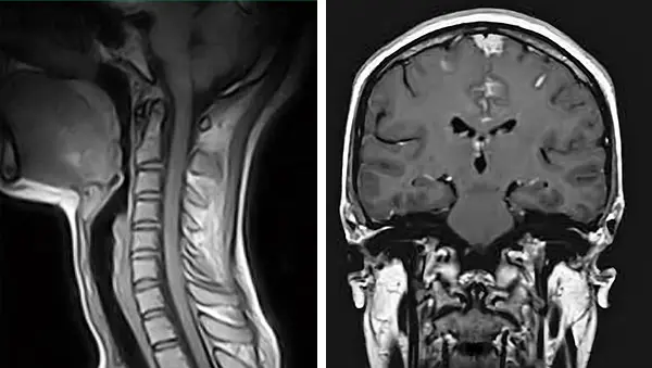
Magnetic resonance imaging (MRI), also known as magnetic resonance tomography (MRT) or tomographic magnetic resonance imaging (MRI), or simply MRI, works on a completely different principle from radiography and does not use X-rays.
The word tomography simply means "layered image"; although MRT contains the word tomography, a tomographic MRI is by no means to be confused with a computed tomography or CT scan. The difference between the 2 is like day and night.
The word tomography simply means "layered image"; although MRT contains the word tomography, a tomographic MRI is by no means to be confused with a computed tomography or CT scan. The difference between the 2 is like day and night.
When the magnetic pulse ceases, depending on the type of tissue and its specific water content, the molecules return to their initial state at different times, which allows the different types of body tissue to be differentiated in the acquired image. This operation is performed in layers and takes a relatively long time, up to 30-40 minutes, depending on how large the area to be scanned is.
While an X-ray or a computed tomography (CT) scan gives a good view of the bones to the exclusion of other tissues, an MRI, on the other hand, allows the soft tissues to be seen rather than the bones. An MRI only allows a two-dimensional view of the scanned area. These two factors make MRI unsuitable for correctly identifying the position of the Atlas in three planes. MRI has the great advantage of being able to produce images without using harmful ionising radiation, but unfortunately it only allows a partial and limited view of what we are interested in seeing.
An MRI does NOT allow the misalignment of the Atlas to be adequately assessed.
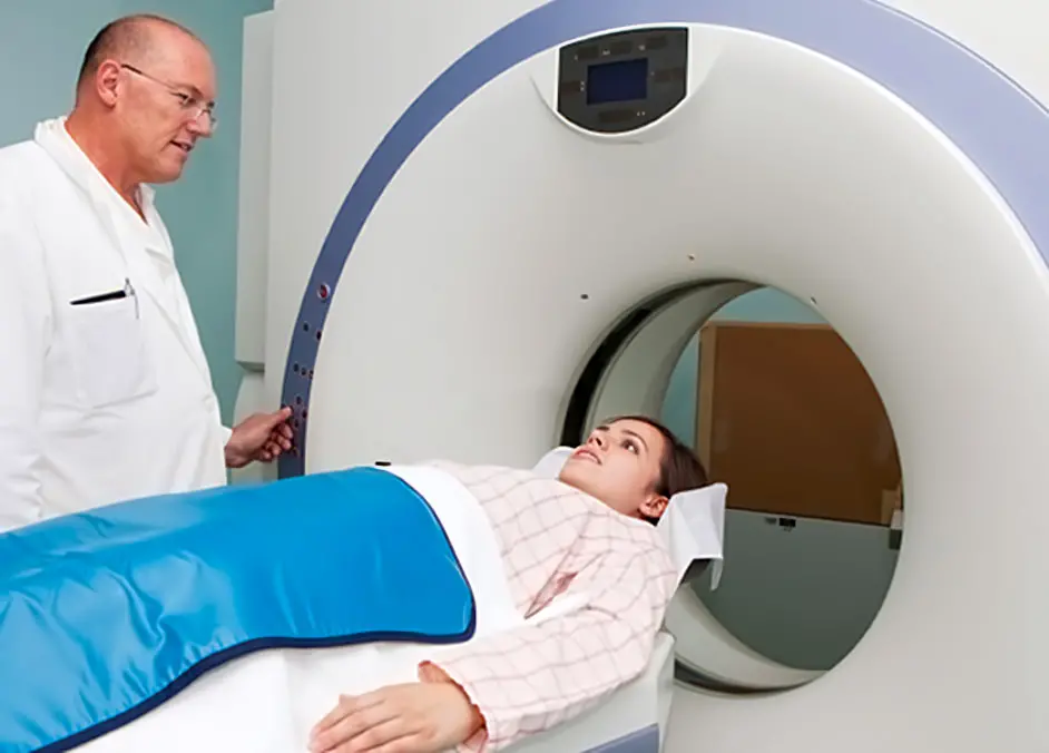
Computed tomography

To put it simply, a computed tomography or CT scan is a machine that takes hundreds of x-rays from multiple perspectives, which are then recombined into a single three-dimensional image using special software.
Modern scanners are all spiral scanners, i.e. the sensor rotates around the subject like a screw, while the couch on which you lie slowly moves forward. The old scanners were called CAT scanners because they only allowed images on one axis.
Modern scanners are all spiral scanners, i.e. the sensor rotates around the subject like a screw, while the couch on which you lie slowly moves forward. The old scanners were called CAT scanners because they only allowed images on one axis.
The radiation of a CT scan of the skull is equivalent to that of 250 chest X-rays, or 2000 microSieverts.
CTCB -Cone Beam Computed Tomography

Cone Beam Computed Tomography is most commonly used in orthodontics. The big advantage of a Cone Beam CT scan over a CT scan is the lower radiation level. Depending on the scanner used, the radiation absorbed ranges from 20 microsieverts in the latest generation of equipment to 120-160 microsieverts in equipment from a few years ago. In the best case, the radiation is therefore 100 times less than that of a spiral CT scan.
In terms of image quality, spiral CT and Cone Beam CT are similar, but Cone Beam has the advantage of producing fewer image artefacts.
However, the lower amount of radiation emitted has the disadvantage of being able to differentiate less between bones than between soft parts, which may be a problem when you want to see the Atlas well. CTCB is usually set up to produce images of the jaw, which is covered by a thin layer of tissue, whereas the Atlas is located much deeper. Increasing the radiation dose solves the problem. The radiation absorbed, however, is many times lower than that of a spiral CT scan.
Cone Beam CT examination is performed in a standing position without having to lie down in a tube (claustrophobia). The advantage is to have the cervical vertebrae under load, avoiding the flattening of the physiological curve of the spine, which invariably occurs when lying down. In this way, it is possible to determine not only vertebral alignment but also the presence of any cervical straightness.
Another factor to take into account is the following: for the orthodontic field, small sensors of the order of 7cm are sufficient. These are too small to capture the entire contour of the skull. To be able to do this, a sensor of at least 15 cm is needed. As explained below, if the entire circumference of the skull is not imaged, the images may be unusable for our purpose.
So:
- A sensor larger than 15 cm is required.
- Radiation must be increased to better differentiate tissues.
It is certainly more difficult to find a suitably equipped centre that can perform a Cone-Beam CT scan than one with a spiral CT scan, as the spiral scanner is much more common. The effort involved in the search is certainly worth the health.

A further advantage of a well performed CT Cone Beam scan is the possibility to see the roots of the teeth and the surrounding bone with a precision not achievable by conventional or panoramic X-rays. In two-dimensional images, multiple molar roots hide each other. With a TCCB, your dentist will be able to discover hidden infections that would otherwise go undetected.
Problems with the oral cavity increase with age and can lead to serious chronic health damage. Take advantage of this opportunity in case you already need a CTCB!
Problems with the oral cavity increase with age and can lead to serious chronic health damage. Take advantage of this opportunity in case you already need a CTCB!
A CT scan as an end in itself, not needed
A CT scan does not provide information on the state of muscle tension and joint function. Therefore, irrespective of the availability of a CT scan, the person must still be expertly assessed manually by an Atlantomed specialist.The CT scan should only be considered as a support for difficult cases. You will therefore understand that it is not sensible and you will not solve the problem by having a CT scan done on your own initiative, in the hope of avoiding a trip. You may have an aligned Atlas, but your neck muscles are so contracted and poorly functional that treatment is necessary and useful. To solve a problem, unlike conventional medicine, we first try the simplest, most sensible and least invasive route, which in our case is functional in 90% of cases. For the remaining 10%, we will take the necessary measures when the time comes. To do otherwise would be absurd.
Rotation of the Atlas or Epistropheus is only one of the parameters that are evaluated as an indication for treatment. Taking this into account, you will understand that it makes absolutely no sense to undergo radiation without first being carefully examined by the Atlas specialist. In most cases, a CT scan is not necessary and should therefore be avoided. There is always time for a CT scan at a later date, should the need arise.
How to perform a CT scan for Atlas misalignment
 In order to assess the upper cervical region correctly, images of the correct anatomical area must first be produced. The ideal portion to be seen includes 1/3 of the skull (lower part) up to C5-C6, if the size of the sensor allows it.
It is not necessary to irradiate the entire skull upwards, nor go too far down the cervical spine. Extending the irradiated anatomical portion, besides being useless for the investigation, subjects the body to unjustified radiation. Going beyond C5-C6 would unnecessarily irradiate the thyroid, an organ that is very sensitive to radiation.
The entire circumference of the skull must be visible on the images. If the front part with the teeth, the back or the side of the skull is missing, the references for a proper evaluation are missing, to the point of making the images unusable.
How to correctly assess a CT scan?
There are circumstances that require a CT scan to be evaluated by the specialist, perhaps because there is a previous one that you had done for other reasons that it would make sense to consult before treatment, or because a new one was done after treatment without success, or even in special cases where malformations or contraindications are present. For this purpose we provide very precise information on how to proceed, without wasting time unnecessarily.In order to see the relative alignments of the vertebrae by means of a CT scan, it is first necessary to reconstruct the volumetric data in a 3D image, then to observe and evaluate the skull from different angles, cutting out the parts that hide useful details. This requires the right software, the right knowledge, knowing a priori what you are looking for and what exactly to look at. So you will understand that it is absolutely useless to show only one image of your CD, as many try to do, no matter what that image is! Without one of the listed measures, the exercise becomes pointless. If we do not receive the file with the volumetric data, but only a few individual images, we cannot do anything!
In order to be able to perform the 3D reconstruction and thus properly assess the cervical spine, we need all the CT volumetric data, i.e. the folder containing the images in DICOM format of about 100-500MB on the CD. All other files are useless and should not be sent. The folder is usually named DICOM, but may also have a different name assigned by the radiologist. If the folder is not at least 50MB, you are NOT sending the RIGHT one! As explained above, it is useless to send X-rays, MRIs and medical or radiographic reports unless explicitly requested for specific reasons. If you have carefully studied and understood what we have written and therefore wish to send us your CT images, the simplest, easiest and quickest way is to send the folder, and only the folder containing the volumetric images, via the service wetransfer.com.
How can I see if my Atlas has shifted?
The radiologist's CD includes a basic utility program that allows you to view the images layer by layer in 2D in the 3 different planes. It may seem obvious to you to be able to detect a misalignment of the Atlas, but what you think you see in the 2D mode is only true under certain very specific conditions.In order to detect a misalignment in the 2D view, a few preliminary steps are necessary. It is impossible to determine the position of the Atlas in relation to the skull if the skull is not previously aligned in its three planes. The head is hardly ever perfectly aligned when it is scanned and this must be taken into account. The question is: what is crooked in relation to what exactly? If the skull on the image is already crooked to begin with, everything you see will also be crooked as a result.
These tricks are systematically ignored and if CT scans are 'badly' done, as is often the case, they become impossible to apply! Looking at the image of a single CT scan, without first making sure that the skull is perfectly aligned on its axes, renders any assessment meaningless.
With 3D image reconstruction, however, some of the gaps in the acquisition can be overcome, with the consequence that the assessment of the relative misalignments becomes much easier and more immediate. However, it is still necessary to rotate the 3D image of the skull several times, on various axes, in order to observe the details useful for the evaluation of misalignment.
A single image, extrapolated from a 3D reconstruction, is in fact useless. I am writing this because many people are used to sending us images of all kinds, asking us for an evaluation. You have to understand that this is an absolutely useless exercise.
Another fact of which you must be well aware: a radiologist is trained to look for specific alterations and structural problems. The misalignment of the Atlas is not one of them. This means that he may not be able to provide a qualified assessment of it. It is very unlikely that you will find any indications of misalignment in the report, and even if you do, they may be wrong. We write this because many people are under the illusion that their vertebrae are fine because they don't read anything specific in the report. "The doctor said everything is fine", which is too bad, because it might not be. This is also why we look at CT scans and evaluate them ourselves, ignoring the reports. We will be happy to show you in detail and share with you what we are able to see and discover in your images.
Conclusions
Once the differences between the various imaging methods are understood, a number of important considerations must be made: Ionising radiation, i.e. X-rays, is not good for your health and should only be used sparingly if you cannot do otherwise and if the risk-benefit ratio is decidedly in favour of the benefit.Exposure to radiation increases the risk of tumours and leukaemia, which is why magnetic resonance imaging is nowadays preferred to CT scans, where appropriate. Unfortunately in the case of the Atlas, as explained in detail above, MRI is not suitable.
Avoid taking unnecessary CT scans lightly!
If the manual evaluation of the vertebrae was not sufficient and it was therefore necessary to resort to a CT scan and thanks to this it was possible to carry out a targeted and effective correction with the subsequent disappearance of the symptoms, then the radiological investigation was justified.
This is not the case if the same result can be achieved by manual examination by the Atlantomed specialist alone. This means that the correct procedure is to visit an Atlantomed centre FIRST in order to evaluate together what to do. If, out of laziness or to save yourself the trip, you intend to do the CT scan beforehand, know that you are not acting in the best way to preserve your future health!
Many people undergo CT examinations with a good dose of recklessness, as if they were going for a walk, without being aware of the long-term consequences of radiation. The radiologist on duty at the time of the examination usually has neither the time nor the foresight to inform you properly. That is why we have explained this topic in detail, so that you can make the most sensible decision.
A CT scan is NOT required to carry out the Atlas correction treatment. Only in certain specific cases, in which the Atlantomed specialist has difficulty in determining the position of the Atlas by manual examination, can the procedure be assessed together.
Important notice
We would like to clarify that the Atlantomed specialist views the images exclusively with the aim of assessing the relative position between the cervical spine and the skull, in order to better understand how to proceed with the relative correction of the vertebrae. It is in no way a medical diagnosis and should not be interpreted as such. Atlas misalignment is not a disease.If you would like a diagnosis of the pathological condition of your spinal column or the state of your tooth roots, please contact a competent radiologist and dentist respectively.
Submitting your MRI scans and medical reports to us in order to obtain a diagnosis is NOT within our competence. Atlantomed is NOT a medical treatment, but a purely mechanical treatment, and for this reason the Atlantomed specialist is in no way intended to replace your doctor.
Read this page carefully: For whom the Atlantomed treatment is intended
Effects on the Axis and other cervical vertebrae

PRIMA
DOPO ATLANTOMED
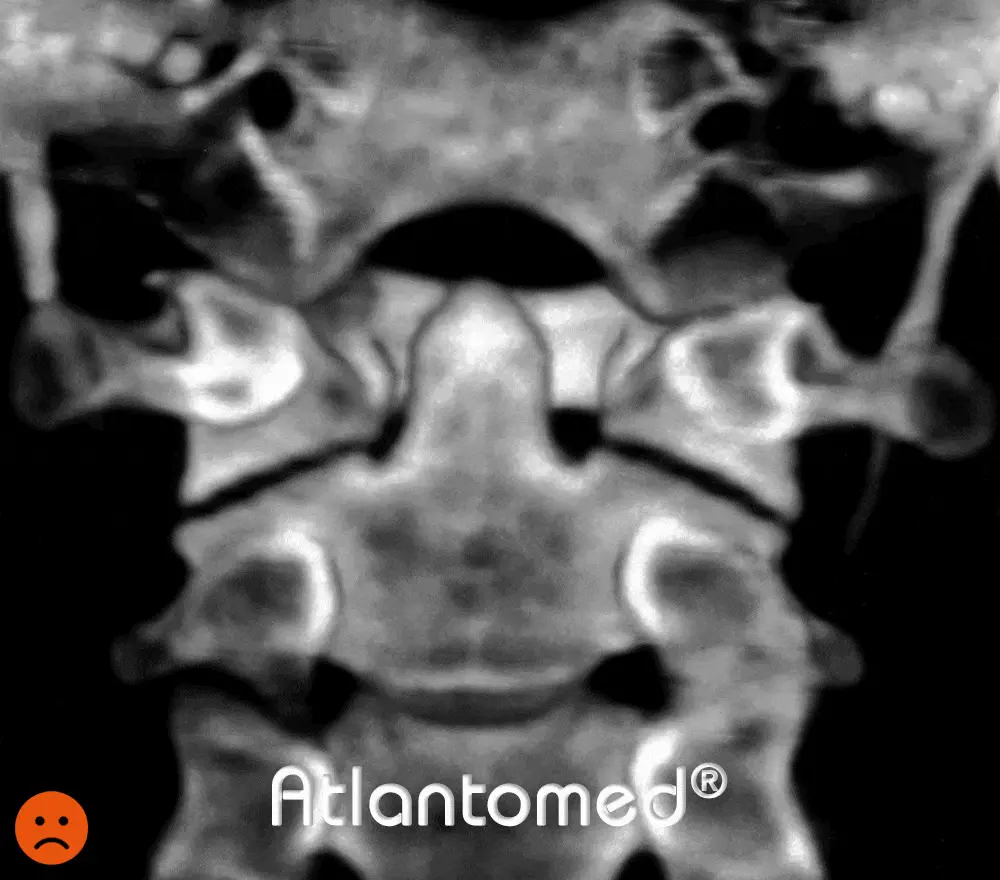
This 3D reconstruction of a spiral CT scan shows a section of the upper cervical vertebrae seen from behind, before and after correction of the Atlas with Atlantomed vibroresonance.
After treatment, the improved alignment of the first Atlas vertebra in relation to the occiput, as well as in relation to the second vertebra, is clearly visible.
The tooth of the epistrophe is more centred in its articulation with the Atlas, while the skull is no longer unbalanced to the right.
After treatment, the improved alignment of the first Atlas vertebra in relation to the occiput, as well as in relation to the second vertebra, is clearly visible.
The tooth of the epistrophe is more centred in its articulation with the Atlas, while the skull is no longer unbalanced to the right.
What people say about us
The only ones with over 9000 testimonials and reviews in several languages! Click to access the related platforms and read many opinions, feedbacks, experiences and testimonials after the Atlantomed vibro-resonance Atlas correction. Be wary of imitations.
Written by: Alfredo Lerro


